previous arrowprevious arrow
medic-176-588-690
medic-176-588-690
Makuladegeneration
Makuladegeneration
old-eye
old-eye
next arrownext arrow

WHAT IS IT ?

Age-related macular degeneration (AMD) is a medical condition which usually affects older adults and results in a loss of vision in the center of the visual field (the macula) because of damage to the retina. It occurs in “dry” and “wet” forms. It is a major cause of blindness and visual impairment in older adults (>50 years). Macular degeneration can make it difficult or impossible to read or recognize faces, although enough peripheral vision remains to allow other activities of daily life.

Picture of the fundus showing intermediate age-related macular degeneration.
Starting from the inside of the eye and going towards the back, the three main layers at the back of the eye are the retina, which contains the nerves; the choroid, which contains the blood supply; and the sclera, which is the white of the eye.

The macula is the central area of the retina, which provides the most detailed central vision.

part-maculapng
Macular Degeneration
macular degeneration

CLASSIFICATION

In the dry (nonexudative) form, cellular debris called drusen accumulate between the retina and the choroid, and the retina can become detached.

In the wet (exudative) form, which is more severe, blood vessels grow up from the choroid behind the retina, and the retina can also become detached.

It can be treated with laser coagulation, and with medication that stops and sometimes reverses the growth of blood vessels. The is a substantial amount of animal model studies showing that stem cells can reverse and or stabilize the vascularization processes, in the retina.

Although some macular dystrophies affecting younger individuals are sometimes referred to as macular degeneration, the term generally refers to age-related macular degeneration (AMD or ARMD).

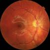
Age-related macular degeneration begins with characteristic yellow deposits (drusen) in the macula, between the retinal pigment epithelium and the underlying choroid. Most people with these early changes (referred to as age-related maculopathy) have good vision. People with drusen can go on to develop advanced AMD. The risk is considerably higher when the drusen are large and numerous and associated with disturbance in the pigmented cell layer under the macula. Recent research suggests that large and soft drusen are related to elevated cholesterol deposits and may respond to cholesterol-lowering agents.

Dry AMD:
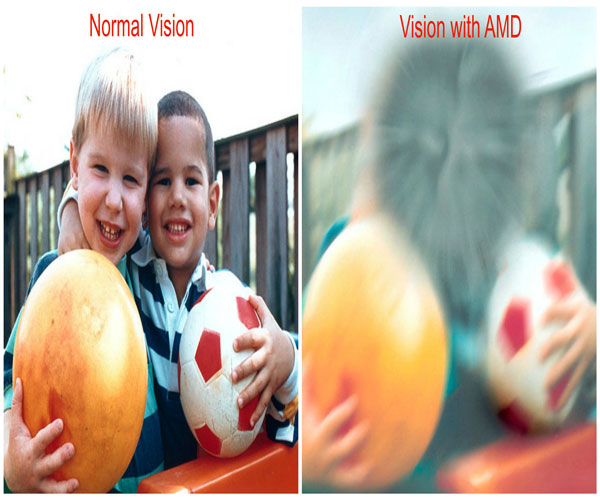
Central geographic atrophy, the “dry” form of advanced AMD, results from atrophy to the retinal pigment epithelial layer below the retina, which causes vision loss through loss of photoreceptors (rods and cones) in the central part of the eye. No medical or surgical treatment is available for this condition, however vitamin supplements with high doses of antioxidants, lutein and zeaxanthin, have been suggested by the National Eye Institute and others to slow the progression of dry macular degeneration and, in some patients, improve visual acuity.

SIGNS AND SYMPTOMS

Macular degeneration by itself will not lead to total blindness. For that matter, only a very small number of people with visual impairment are totally blind. In almost all cases, some vision remains. Other complicating conditions may possibly lead to such an acute condition (severe stroke or trauma, untreated glaucoma, etc.), but few macular degeneration patients experience total visual loss.

The area of the macula comprises only about 2.1% of the retina, and the remaining 97.9% (the peripheral field) remains unaffected by the disease. Interestingly, even though the macula provides such a small fraction of the visual field, almost half of the visual cortex is devoted to processing macular information.

The loss of central vision profoundly affects visual functioning. It is not possible, for example, to read without central vision. Pictures that attempt to depict the central visual loss of macular degeneration with a black spot do not really do justice to the devastating nature of the visual loss. This can be demonstrated by printing letters 6 inches high on a piece of paper and attempting to identify them while looking straight ahead and holding the paper slightly to the side. Most people find this difficult to do.

There is a loss of contrast sensitivity, so that contours, shadows, and color vision are less vivid. The loss in contrast sensitivity can be quickly and easily measured by a contrast sensitivity test performed either at home or by an eye specialist.

Macular Degeneration

CAUSES

The practical application of AMD-associated markers, such as seen above, is in the prediction of progression of AMD from early stages of the disease to neovascularization.

A family of immune mediators has been shown to be plentiful in drusen, the cellular debris associated with macular degeneration. Complement factor H (CFH) is an important inhibitor of this inflammatory cascade and a disease-associated polymorphism in the CFH gene strongly associates with AMD. Thus an AMD pathophysiological model of chronic low grade complement activation and inflammation in the macula has been advanced. Lending credibility to this has been the discovery of disease-associated genetic polymorphisms in other elements of the complement cascade including complement component 3 (C3).

The role of retinal oxidative stress in the etiology of AMD by causing further inflammation of the macula is suggested by the enhanced rate of disease in smokers and those exposed to UV irradiation. Mitochondria are a major source of oxygen free radicals that occur as a byproduct of energy metabolism. Mitochondrial gene polymorphisms, such as that in the MT-ND2 molecule, predicts wet AMD.

Aging: Approximately 10% of patients 66 to 74 years of age will have findings of macular degeneration. The prevalence increases to 30% in patients 75 to 85 years of age.

Family history: The lifetime risk of developing late-stage macular degeneration is 50% for people that have a relative with macular degeneration, versus 12% for people that do not have relatives with macular degeneration, a fourfold higher risk. (similar lifestyles and of course genetics)
Macular degeneration gene: The genes for the complement system proteins factor H (CFH), factor B (CFB) and factor 3 (C3) have been determined to be strongly associated with a person’s risk for developing macular degeneration.

Mutation of the ATP synthase gene: Retinitis pigmentosa (RP) is a genetically linked dysfunction of the retina and is related to mutation of the adenosine triphosphate (ATP) synthase gene 615.1617
Stargardt’s disease (STGD), also known as juvenile macular degeneration) is an autosomal recessive retinal disorder characterized by a juvenile-onset macular dystrophy, alterations of the peripheral retina, and subretinal deposition of lipofuscin-like material.

Drusen: CMSD studies indicate that drusen are similar in molecular composition to plaques and deposits in other age-related diseases such as Alzheimer’s disease and atherosclerosis.

Arg80Gly variant of the complement protein C3.

Cardiovascular status: High cholesterol, obesity.

High fat intake is associated with an increased risk of macular degeneration in both women and men.

Oxidative stress: It has been proposed that age-related accumulation of low-molecular-weight, phototoxic, pro-oxidant melanin oligomers within lysosomes in the retinal pigment epithelium may be partly responsible for decreasing the digestive rate of photoreceptor outer rod segments (POS) by the RPE.

Fibulin-5 mutation: Rare forms of the disease are caused by geneic defects in fibulin-5, in an autosomal dominant manner.

Exposure to sunlight especially blue light: There is conflicting evidence as to whether exposure to sunlight contributes to the development of macular degeneration.

Smoking: Smoking tobacco increases the risk of macular degeneration by two to three times that of someone who has never smoked, and may be the most important modifiable factor in its prevention.

Deletion of CFHR3 and CFHR1: Deletion of the complement factor H-related genes CFHR3 and CFHR1 protects against age-related macular degeneration.

Hypertension: Also known as high blood pressure.

GENETIC TESTING

A powerful predictor of AMD is found on chromosome 10q26 at LOC 387715. An insertion/deletion polymorphism at this site reduces expression of the ARMS2 gene though destabilization of its mRNA through deletion of the polyadenylation signal. ARMS2 protein may localize to the mitochondria and participate in energy metabolism, though much remains to be discovered about its function.

Other gene markers of progression risk includes Tissue Inhibitor of Metalloproteinase 3 (TIMP3) suggesting a role for intracellular matrix metabolism in AMD progression. Variations in cholesterol metabolising genes such as the hepatic lipase (LIPC), cholesterol ester transferase (CETP), lipoprotein lipase (LPL) and the ABC-binding cassette A1 (ABCA1) correlate with disease progression, Early stigmata of disease, drusen, are rich in cholesterol, offering face validity to the results of genome wide association studies

pngeye

Macular-Degeneration-Photo

TREATMENT

The newest finding confirm that immune mediators of the complement cascade are one of the responsible mechanisms creating this disorder. Immune modulation has been the hallmark of stem cell therapy and is well researched in a number of other diseases. The second mechanism of decreasing inflammation commonly seen post stem cell therapy, is also responsible for the decrease in macular degeneration.

Although there is a limited amount of clinical or published data, considering the very low risk from both the procedure and the use of autologous cells this approach has significant merit. A number of new clinical trials have been approved based on the genetic and immune markers expressions and the active input that stem cells can affect on these pathways.
Unfortunately the studies just announced are for embryonic cell use and will take years to translate to clinical practice. We feel strongly that autologous cells have shown similar potentials, have a high safety index and can be used currently with good to excellent potential outcomes.

Additional medical interventions targeting the lipids (fats) along with the use of antioxidant therapy should maximize the impact of the cellular therapy and result in stabilization or even partial reversal of macular degeneration.

Antioxidant therapy is somewhat controversial due to contradictory results of a number of studies and the conflict of interest suggested by some. The different results may be more of a effect of lack of superior products and proper testing of the patients, prior to supplementation. I believe that most findings suggest strongly that the use of the antioxidants can be a positive input and should be considered for the appropriate patient.

Over the last 12 years, World Stem Cell Clinic has preformed  Intraocular injections (also known as “intravitreal injections” and “eye injections”) for treatment of many retinal diseases. An intravitreal injection is a procedure performed to introduce autologous stem cells from bone marrow, adipose, peripheral blood or allogenic cells from amnion, cord blood, wj, plasma proteins directly into (“intra-“) the gel-filled center of your eye (‘-vitreal”) Our proprietary protocol and process based on each unique patient status.

After a review of your medical records and discussions with medical staff, a protocol is designed especially for you. Specifics of your condition are addressed along with any special needs. It may be similar to the one illustrated below:

  • DAY 1

    At the clinic you will be examined by our physicians. Information including any risks and expectations concerning your treatment, plus answers to any questions you may have will be addressed. A blood draw, to determine chemistries prior to the treatment will be collected and any medication required may be administered. Then you will return to your hotel for a restful day or a good nights sleep.

  • DAY 2

    Our physician’s will review the laboratory results, determine if the results are within range, and discuss the response with you. They may or may not provide additional medications and may add adjunctive treatments. They will determine if you would return to the hotel, with little restriction on your activities, or possibly go forward with with your treatment that day.

  • DAY 3

    If either a bone marrow or adipose collection will be utilized and the cell count / viability is appropriate for harvest. We typically use local anaesthetics for adults and general anaesthesia for youngsters. The entire procedure normally takes less than 30 minutes. Although some pain is felt when the needle is inserted, most patients do not find the bone marrow collection procedure particularly painful or uncomfortable.
    We recently placed a number of videos on our website interviewing our patient’s who discuss the procedure and their lack of discomfort.
    After the collection you may return to the hotel, with some restrictions. The bone marrow or adipose collected is processed in our contract State-Of-Art laboratory by trained staff, under the supervision of the lab physician.
    As an alternative to the above, umbilical cord derived calls, plasma, protein and extracellular vesicles based on your unique treatment needs may be used based on the patient’s individual medical condition and options.

  • DAY 4

    You will be treated by an intrabulbar (into the eye) injection and IV infusion by one of our board certified othalmologist. This allows the stem cells to begin activity in the affected areas directly. When the procedure is performed, you will be given a very mild anaesthetic into the eye and be asked to restrict your activities for that day.

  • DAY 5

    You will receive a post-treatment examination and evaluation prior to release. Return home or optionally there may be the use of additional ancillary therapies to enhance the procedure and additional evaluations.

WHAT MAKES OUR TREATMENT DIFFERENT

  • At World Stem Cell Clinic and The Royal British Medical Center we practice “Patient Precision Medicine (PPM)” which is a treatment model that proposes the customization of the treatment to each unique patient based on their medical history, stage of disease, exam results, time available for treatment and a patient orientation meeting with our Doctors before determining the best treatment for each unique patient.


  • Our staff physicians are all board certified, in their field with years of experience. Your team includes both primary and ancillary care professionals devoted to maximizing your benefits from the procedures. We enroll you in an open registry to track your changes independently, for up to 5 years.


  • To maintain our “Patient Precision Medicine (PPM)” services for you we may use peripheral blood, bone marrow, adipose or umbilical cord derived cells, plasma, proteins and Extracellular Vesicles based your unique treatment needs with mutual agreement.


  • As our patient we also keep you abreast of the newest developments in treatment research. This is an ongoing relationship to maintain and enhance your health.


  • Our promise is to provide you with travel and lodging support, access to bilingual staff members throughout the entire process and most importantly the best medical care possible.

Online Consult Form

We will contact you within one business day.


ADVERTISEMENT FOR: 
WORLD STEM CELL CLINIC LLC

THE ROYAL BRITISH MEDICAL CENTER

Do you qualify for a treatment?
REQUEST FREE CONSULTATION!

* indicates required field