previous arrowprevious arrow
Blockedslider
Blockedslider
Peripheral-Arterial-Disease-slider
next arrownext arrow

WHAT IS IT ?

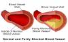
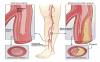
Peripheral vascular disease (PVD), commonly referred to as peripheral arterial disease (PAD) or peripheral artery occlusive disease (PAOD), refers to the obstruction of large arteries not within the coronary, aortic arch vasculature, or brain. PVD can result from atherosclerosis, inflammatory processes leading to stenosis, an embolism, or thrombus formation. It causes either acute or chronic ischemia (lack of blood supply). Often peripheral arterial disease(PAD) is a term used to refer to atherosclerotic blockages found in the lower extremity.

PVD also includes a subset of diseases classified as microvascular diseases resulting from episodal narrowing of the arteries (Raynaud’s phenomenon), or widening thereof (erythromelalgia), i.e. vascular spasms.

Levels of disability are divided into 6 subsets. The classification system, by Rutherford, consists of three grades and six categories:
Mild claudication
Moderate claudication
Severe claudication
Ischemic pain at rest
Minor tissue loss
Major tissue loss
Symptoms
About 20% of patients with mild peripheral arterial disease(PAD) may be asymptomatic; other symptoms include:[1]
Claudication – pain, weakness, numbness, or cramping in muscles due to decreased blood flow,Sores, wounds, or ulcers that heal slowly or not at all.

Noticeable change in color (blueness or paleness) or temperature (coolness) when compared to the other limb
Diminished hair and nail growth on affected limb and digits.

Peripheral Arterial Disease Treatments
Peripheral Arterial Disease Treatment

SIGNS AND SYMPTOMS

Bouchard’s nodes and Heberden’s nodes may form in osteoarthritis
The main symptom is pain, causing loss of ability and often stiffness. “Pain” is generally described as a sharp ache, or a burning sensation in the associate muscles and tendons. OA can cause a crackling noise (called “crepitus”) when the affected joint is moved or touched, and patients may experience muscle spasm and contractions in the tendons. Occasionally, the joints may also be filled with fluid. Humid and cold weather increases the pain in many patients.

OA commonly affects the hands, feet, spine, and the large weight bearing joints, such as the hips and knees, although in theory, any joint in the body can be affected. As OA progresses, the affected joints appear larger, are stiff and painful, and usually feel better with gentle use but worse with excessive or prolonged use, thus distinguishing it from rheumatoid arthritis.

In smaller joints, such as at the fingers, hard bony enlargements, called Heberden’s nodes (on the distal interphalangeal joints) and/or Bouchard’s nodes (on the proximal interphalangeal joints), may form, and though they are not necessarily painful, they do limit the movement of the fingers significantly. OA at the toes leads to the formation of bunions, rendering them red or swollen. Some people notice these physical changes before they experience any pain.OA is the most common cause of joint effusion, sometimes called water on the knee in lay terms, an accumulation of excess fluid in or around the knee joint.

CAUSES

Risk factors contributing to peripheral arterial disease(PAD) are the same as those for therosclerosis:
Smoking – tobacco use in any form is the single most important modifiable cause of PVD internationally. Smokers have up to a tenfold increase in relative risk for PVD in a dose-related effect. Exposure to second-hand smoke from environmental exposure has also been shown to promote changes in blood vessel lining (endothelium) which is a precursor to atherosclerosis.

Diabetes mellitus – causes between two and four times increased risk of PVD by causing endothelial and smooth muscle cell dysfunction in peripheral arteries. Diabetics account for up to 70% of nontraumatic amputations performed, and a known diabetic who smokes runs an approximately 30% risk of amputation within 5 years.

Dyslipidemia (high low density lipoprotein [LDL] cholesterol, low high density lipoprotein [HDL] cholesterol) – elevation of total cholesterol, LDL cholesterol, and triglyceride levels each have been correlated with accelerated peripheral arterial disease(PAD). Correction of dyslipidemia by diet and/or medication is associated with a major improvement in short-term rates of heart attack and stroke This benefit is gained even though current evidence does not demonstrate a major reversal of peripheral and/or coronary atherosclerosis.
Hypertension – elevated blood pressure is correlated with an increase in the risk of developing peripheral arterial disease(PAD), as well as in associated coronary and cerebrovascular events (heart attack and stroke).

Risk of peripheral arterial disease(PAD) also increases in individuals who are over the age of 50, male, obese, or with a family history of vascular disease, heart attack, or stroke.Other risk factors, which are being studied, include levels of various inflammatory mediators such as C-reactive protein, homocysteine.

artery
PAD

DIAGNOSIS

Upon suspicion of PVD, the first-line study is the ankle brachial pressure index (ABPI/ABI). This non-invasive and cost effect test is easily done in your physicians office. When the blood pressure readings in the ankles are lower than that in the arms, blockages in the arteries, which provide blood from the heart to the ankle, are suspected. An ABI ratio less than 0.9 is consistent with PVD; values of ABI below 0.8 indicate moderate disease and below 0.4 imply severe ischemic disease.[1]

It is possible for conditions which stiffen the vessel walls (such as calcifications that occur in the setting of chronic diabetes) to produce false negatives usually, but not always, indicated by abnormally high ABIs (> 1.3). Such results and suspicions require further investigation using ultrasound to fully evaluate.

If your ABIs are abnormal typically a lower limb doppler ultrasound examination to look at site and extent of atherosclerosis should be ordered.? Other imaging can be performed by angiography,[1] where a catheter is inserted into the common femoral artery and selectively guided to the artery in question. While injecting a radiodense contrast agent an X-ray is taken. Any flow limiting stenoses found in the x-ray can be identified and potentially treated by atherectomy, angioplasty or stenting, however these procedure are not risk free.

Modern multislice computerized tomography (CT) scanners provide direct imaging of the arterial system as an alternative to angiography. CT provides complete evaluation of the aorta and lower limb arteries without the need for an angiogram’s arterial injection of contrast agent. This method employs a significant exposure to radiation and one should always use NAC, a supplement if contrast is used.

peripheral arterial disease(PAD)

TREATMENT

It should be noted that individuals with peripheral arterial disease(PAD) have an “exceptionally elevated risk for cardiovascular events and the majority will eventually die of a cardiac or cerebrovascular etiology”;There is a direct prognosis correlated with the severity of the peripheral arterial disease(PAD) as measured by the Ankle brachial pressure index (ABPI). If the large-vessels are involved with peripheral arterial disease(PAD) , this increases mortality from cardiovascular disease significantly. peripheral arterial disease(PAD) carries a greater than “20% risk of a coronary event in 10 years”.

There has been a tremendous amount of work done it the area of peripheral arterial disease(PAD) treatment due to the staggering costs and disability associated with this growing population of patients. The overwhelming costs for the disability, even with minimal expression of symptoms, confounds and taxes the health care system. The use of a minimally invasive therapy, with limited risk, is a welcome addition for any patients.

Unfortunately the majority of patients will never be offered this limb saving and quality of life preserving therapy. The current regulation of medical care of the FDA have not approved the use of autologous stem cells from bone marrow, adipose, peripheral blood or allogenic cells from amnion, cord blood, wj, plasma proteins therapy in the State and Canada. The good news is that by travelling outside of the country you can receive this therapeutic intervention. SAVE YOUR LIMB!

After a review of your medical records and discussions with medical staff, a protocol is designed especially for you. Specifics of your condition are addressed along with any special needs. It may be similar to the one illustrated below:

  • DAY 1

    At the clinic you will be examined by our physicians. Information including any risks and expectations concerning your treatment, plus answers to any questions you may have will be addressed. A blood draw, to determine chemistries prior to the treatment will be collected and any medication required may be administered. Then you will return to your hotel for a restful day or a good nights sleep.

  • DAY 2

    Our physician’s will review the laboratory results, determine if the results are within range, and discuss the response with you. They may or may not provide additional medications and may add adjunctive treatments. They will determine if you would return to the hotel, with little restriction on your activities, or possibly go forward with with your treatment that day.

  • DAY 3

    If either a bone marrow or adipose collection will be utilized and the cell count / viability is appropriate for harvest. We typically use local anaesthetics for adults and general anaesthesia for youngsters. The entire procedure normally takes less than 30 minutes. Although some pain is felt when the needle is inserted, most patients do not find the bone marrow collection procedure particularly painful or uncomfortable.
    We recently placed a number of videos on our website interviewing our patient’s who discuss the procedure and their lack of discomfort.
    After the collection you may return to the hotel, with some restrictions. The bone marrow or adipose collected is processed in our contract State-Of-Art laboratory by trained staff, under the supervision of the lab physician.
    As an alternative to the above, umbilical cord derived calls, plasma, protein and extracellular vesicles based on your unique treatment needs may be used based on the patient’s individual medical condition and options.

  • DAY 4

    You will be treated with both an IV infusion and injections, into the lower limb. This route transports the cells into the local tissue and the general circulation. The procedure is minimally uncomfortable and takes ~1.5 hours.
    You will be required to restrict activities and spend a restfully day at the hotel.

  • DAY 5

    At the clinic you will receive a post-treatment examination and evaluation. Return home or optionally there may be the use of additional ancillary therapies to enhance the procedure.

WHAT MAKES OUR TREATMENT DIFFERENT

  • At World Stem Cell Clinic and The Royal British Medical Center we practice “Patient Precision Medicine (PPM)” which is a treatment model that proposes the customization of the treatment to each unique patient based on their medical history, stage of disease, exam results, time available for treatment and a patient orientation meeting with our Doctors before determining the best treatment for each unique patient.


  • Our staff physicians are all board certified, in their field with years of experience. Your team includes both primary and ancillary care professionals devoted to maximizing your benefits from the procedures. We enroll you in an open registry to track your changes independently, for up to 5 years.


  • To maintain our “Patient Precision Medicine (PPM)” services for you we may use peripheral blood, bone marrow, adipose or umbilical cord derived cells, plasma, proteins and Extracellular Vesicles based your unique treatment needs with mutual agreement.


  • As our patient we also keep you abreast of the newest developments in treatment research. This is an ongoing relationship to maintain and enhance your health.


  • Our promise is to provide you with travel and lodging support, access to bilingual staff members throughout the entire process and most importantly the best medical care possible.


ADVERTISEMENT FOR: 
WORLD STEM CELL CLINIC LLC

THE ROYAL BRITISH MEDICAL CENTER

Do you qualify for a treatment?
REQUEST FREE CONSULTATION!

* indicates required field