previous arrow
osteo1
osteo1
pm-0216-med-img_8294-921
next arrow

WHAT IS IT ?

Rheumatoid arthritis (RA) Osteoarthritis (OA)

Osteoarthritis (OA) also known as degenerative arthritis or degenerative joint disease or osteoarthrosis, is a group of mechanical abnormalities involving degradation of joints,including articular cartilage and subchondral bone. Symptoms may include joint pain, tenderness, stiffness, locking, and sometimes an effusion. A variety of causes—hereditary, developmental, metabolic, and mechanical—may initiate processes leading to loss of cartilage. When bone surfaces become less well protected by cartilage, bone may be exposed and damaged. As a result of decreased movement secondary to pain, regional muscles may atrophy, and ligaments may become more lax. Treatment generally involves a combination of exercise, lifestyle modification, and analgesics. If pain becomes debilitating, joint replacement surgery may be used to improve the quality of life. OA is the most common form of arthritis, and the leading cause of chronic disability in the United States. It affects about 8 million people in the United Kingdom and nearly 27 million people in the United States.

Rheumatoid arthritis (RA) is a long-term autoimmune disorder that primarily affects joints. It typically results in warm, swollen, and painful joints. Pain and stiffness often worsen following rest. Most commonly, the wrist and hands are involved, with the same joints typically involved on both sides of the body. The disease may also affect other parts of the body. This may result in a low red blood cell countinflammation around the lungs, and inflammation around the heart. Fever and low energy may also be present. Often, symptoms come on gradually over weeks to months.

Rheumatoid Arthritis (RA) Osteoarthritis (OA)
osteoarthrosis treatment

SIGNS AND SYMPTOMS

Rheumatoid arthritis (RA) Osteoarthritis (OA)

Osteoarthritis (OA) Bouchard’s nodes and Heberden’s nodes may form in osteoarthritis.The main symptom is pain, causing loss of ability and often stiffness. “Pain” is generally described as a sharp ache, or a burning sensation in the associate muscles and tendons. OA can cause a crackling noise (called “crepitus”) when the affected joint is moved or touched, and patients may experience muscle spasm and contractions in the tendons. Occasionally, the joints may also be filled with fluid. Humid and cold weather increases the pain in many patients.
OA commonly affects the hands, feet, spine, and the large weight bearing joints, such as the hips and knees, although in theory, any joint in the body can be affected. As OA progresses, the affected joints appear larger, are stiff and painful, and usually feel better with gentle use but worse with excessive or prolonged use, thus distinguishing it from rheumatoid arthritis.
In smaller joints, such as at the fingers, hard bony enlargements, called Heberden’s nodes (on the distal interphalangeal joints) and/or Bouchard’s nodes (on the proximal interphalangeal joints), may form, and though they are not necessarily painful, they do limit the movement of the fingers significantly. OA at the toes leads to the formation of bunions, rendering them red or swollen. Some people notice these physical changes before they experience any pain.
OA is the most common cause of joint effusion, sometimes called water on the knee in lay terms, an accumulation of excess fluid in or around the knee joint.

Rheumatoid arthritis (RA) primarily affects joints, but it also affects other organs in more than 15–25% of individuals.  Arthritis of joints involves inflammation of the synovial membrane. Joints become swollen, tender and warm, and stiffness limits their movement. With time, multiple joints are affected (polyarthritis). Most commonly involved are the small joints of the hands, feet and cervical spine, but larger joints like the shoulder and knee can also be involved.  Synovitis can lead to tethering of tissue with loss of movement and erosion of the joint surface causing deformity and loss of function.

RA typically manifests with signs of inflammation, with the affected joints being swollen, warm, painful and stiff, particularly early in the morning on waking or following prolonged inactivity. Increased stiffness early in the morning is often a prominent feature of the disease and typically lasts for more than an hour. Gentle movements may relieve symptoms in early stages of the disease. These signs help distinguish rheumatoid from non-inflammatory problems of the joints, such as osteoarthritis. In arthritis of non-inflammatory causes, signs of inflammation and early morning stiffness are less prominent with stiffness typically less than one hour, and movements induce pain caused by mechanical arthritis.[16] The pain associated with RA is induced at the site of inflammation and classified as nociceptive as opposed to neuropathic. The joints are often affected in a fairly symmetrical fashion, although this is not specific, and the initial presentation may be asymmetrical.

CAUSES

Rheumatoid arthritis (RA) Osteoarthritis (OA)

Osteoarthritis (OA) Some investigators believe that mechanical stress on joints underlies all osteoarthritis, with many and varied sources of mechanical stress, including misalignments of bones caused by congenital or pathogenic causes; mechanical injury; overweight; loss of strength in muscles supporting joints; and impairment of peripheral nerves, leading to sudden or uncoordinated movements that overstress joints. However exercise, including running in the absence of injury, has not been found to increase one’s risk of developing osteoarthritis. Nor has cracking ones knuckles been found to play a role.

Rheumatoid arthritis (RA) is a chronic autoimmune disorder the causes of which are not completely understood. It is a systemic (whole body) disorder principally affecting synovial tissues. There is no evidence that physical and emotional effects or stress could be a trigger for the disease. The many negative findings suggest that either the trigger varies, or that it might, in fact, be a chance event inherent with the immune response.

A family history of RA increases the risk around three to five times; as of 2017 it was estimated that genetics may account for between 40 and 65% of cases of seropositive RA, but only around 20% for seronegative RA.  RA is strongly associated with genes of the inherited tissue type major histocompatibility complex (MHC) antigen HLA-DR4 is the major genetic factor implicated – but its relative importance varies across ethnic groups.

osteoarthrosis treatment
Rheumatoid Arthritis (RA) Osteoarthritis (OA)

DIAGNOSIS

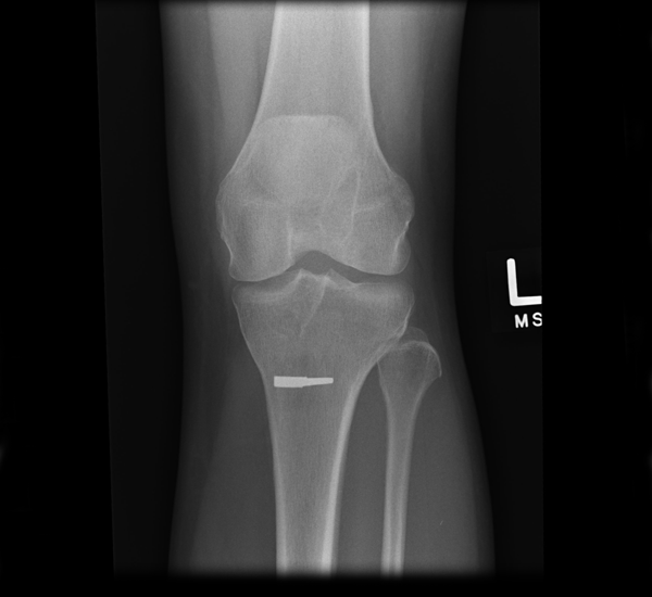
Diagnosis is made with reasonable certainty based on history and clinical examination. X-rays may confirm the diagnosis. The typical changes seen on X-ray include: joint space narrowing, subchondral sclerosis (increased bone formation around the joint), subchondral cyst formation, and osteophytes. Plain films may not correlate with the findings on physical examination or with the degree of pain. Usually other imaging techniques are not necessary to clinically diagnose osteoarthritis.
In 1990, the American College of Rheumatology, using data from a multi-center study, developed a set of criteria for the diagnosis of hand osteoarthritis based on hard tissue enlargement and swelling of certain joints. These criteria were found to be 92% sensitive and 98% specific for hand osteoarthritis versus other entities such as rheumatoid arthritis and spondyloarthropathies.
Related pathologies whose names may be confused with osteoarthritis include pseudo-arthrosis. This is derived from the Greek words pseudo, meaning “false”, and arthrosis, meaning “joint.” Radiographic diagnosis results in diagnosis of a fracture within a joint, which is not to be confused with osteoarthritis which is a degenerative pathology affecting a high incidence of distal phalangeal joints of female patients.

TREATMENT

Rheumatoid arthritis (RA) Osteoarthritis (OA)

The large and expanding body of publications utilizing stem cell technology in orthopedic applications indicates that the infusion of stem cells and growth factors result in the modulation of T cell activity, decreased inflammatory chemicals and the stimulation of the chondocytes.
This combination of responses is probably the basis for the results seen in the referenced clinical trials. There clearly needs to be an increased utilization of the stem cell approach to safely address this condition. Unfortunately this is unlikely, as the use of expensive and risky pharmaceutical agents has taken the forefront. The limited options for this disorder suggest other avenues of treatment should preclude the surgeries that typical mark the end point of the disease process.
We at World Stem Cell clinic feel strongly that the non-responders to conventional therapies should utilize autologous stem cells from bone marrow, adipose, peripheral blood or allogenic cells from amnion, cord blood, wj, plasma proteins approach, prior to the use of the surgical and/or long-term steroidal or even non-steroidal medicinal applications. We have had great success with Osteoarthritis (OA) and Rheumatoid arthritis (RA) allowing patient to have a better quality of life and retard the advance and pain of the disease.

NOTE: We found that the  BMAC  procedure was less effective than the hUCB-MSC procedure for cartilage regeneration in medial unicompartmental knee OA even though the clinical outcomes improved regardless of which treatment was administered.

After a review of your medical records and discussions with medical staff, a protocol is designed especially for you. Specifics of your condition are addressed along with any special needs. It may be similar to the one illustrated below:

  • DAY 1

    At the clinic you will be examined by our physicians. Information including any risks and expectations concerning your treatment, plus answers to any questions you may have will be addressed. A blood draw, to determine cell counts and other chemistries will be collected and cell expansion medication may be administered. Then you will return to your hotel for a restful day or a good nights sleep.

  • DAY 2

    Our physician’s will review the laboratory results, determine if the cell count is within range, and discuss the response to the stimulation. They may or may not provide additional cell expansion medications and may add adjunctive treatments. The levels of your response will determine if you would return to the hotel, with little restriction on your activities, or possibly go forward with harvesting and processing your cells.

  • DAY 3

    Depending on your Exam we will utilize autologous stem cells from bone marrow, adipose, peripheral blood or allogenic cells from amnion, cord blood, wj, plasma proteins.

    We typically use local anaesthetics for adults and general anaesthesia for youngsters. The entire procedure normally takes less than 30 minutes. Although some pain is felt when the needle is inserted, most patients do not find the bone marrow collection procedure particularly painful or uncomfortable.
    We recently placed a number of videos on our website interviewing our patient’s who discuss the procedure and their lack of discomfort.
    After the collection you may return to the hotel, with some restrictions. The bone marrow or adipose collected is processed in our contract State-Of-Art laboratory by trained staff, under the supervision of the lab physician.
    As an alternative to the above, umbilical cord derived calls, plasma, protein and extracellular vesicles based on your unique treatment needs may be used based on the patient’s individual medical condition and options.

  • DAY 4

    Cellular re-implantation if not done on day 3. This will require a restful day and observation, including a post treatment examination. Additional physical or occupational therapy per the individual protocol.

  • DAY 5

    Meet with physician/s to reassess your condition and discuss your improvements and provide suggestions on what you can do to maximize your recovery and what not to do. We will follow up with you in 30days, 90days and 180 days to evaluate your progress.
    After consultation you may return home or optionally there may be the use of additional ancillary therapies to enhance the procedure prior to leaving.

WHAT MAKES OUR TREATMENT DIFFERENT

  • At World Stem Cell Clinic and The Royal British Medical Center we practice “Patient Precision Medicine (PPM)” which is a treatment model that proposes the customization of the treatment to each unique patient based on their medical history, stage of disease, exam results, time available for treatment and a patient orientation meeting with our Doctors before determining the best treatment for each unique patient.


  • Our staff physicians are all board certified, in their field with years of experience. Your team includes both primary and ancillary care professionals devoted to maximizing your benefits from the procedures. We enroll you in an open registry to track your changes independently, for up to 5 years.


  • To maintain our “Patient Precision Medicine (PPM)” services for you we may use peripheral blood, bone marrow, adipose or umbilical cord derived cells, plasma, proteins and Extracellular Vesicles based your unique treatment needs with mutual agreement.


  • As our patient we also keep you abreast of the newest developments in treatment research. This is an ongoing relationship to maintain and enhance your health.


  • Our promise is to provide you with travel and lodging support, access to bilingual staff members throughout the entire process and most importantly the best medical care possible.


ADVERTISEMENT FOR: 
WORLD STEM CELL CLINIC LLC

THE ROYAL BRITISH MEDICAL CENTER

Do you qualify for a treatment?
REQUEST FREE CONSULTATION!

* indicates required field